![]()
A Rapid Image Acquisition System and Method for Focus Stacking Photomontage in Microscopy
Modern image presentation benefits greatly from the use of well-known focus stacking photomontage, i.e., the taking of a plurality of images at successive focal distances, followed by mathematical processing and combining of the images to produce a resultant single image with an extended depth of field (DOF). This is useful when viewing subjects whose visible features lie at various depths greater than the depth of field of an objective lens in use, and also objects that are tilted or have irregular surfaces. In the past, images to be included in a stack were taken one-at-a-time. A first image was taken, then the focus of a camera or the position of a subject was changed and a second image was taken, and so forth. The images were then submitted to a software program for processing and focus stacking into a final result. This process works well, however it takes time. We have developed a system and method for taking a plurality of images at video rates, i.e. at many tens of images per second, without the need for moving a subject or manually refocusing a camera between shots. Our system greatly increases speed, convenience, and workflow in obtaining images with an extended depth of field. Moving subjects and panning of subjects are possible. There are many new possibilities with our new photomontage system. A detailed description of our system is contained in our U.S. patent 8,212,915 and in white papers at: Relay Lens and Video Photomontage. Our system also applies to cameras, telescopes, binoculars, and monoculars. Some examples of images taken using our photomontage system are shown below. More examples will be added periodically so be sure to check back. |
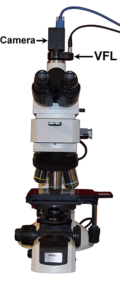
| How it works: In one version of our system, a variable focus lens (VFL) is inserted in the trinocular port of a microscope and focuses an image of a specimen onto an image sensor. A control unit adjusts the focus of the variable focus lens with each image taken by the sensor. A series of images, each at a different focal depth, are processed by focus stacking software. After processing, an all-in-focus image is delivered to a display and storage unit for viewing and saving. |
| Videos: | |
 |
Time lapse: Free-ranging amoeba is able to move horizontally and vertically in a concave microscope slide, not squeezed between a slide and a coverslip. Each image was distilled from a stack of 10 individual images taken at a rate of 60 frames per second. |
 |
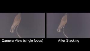
Time lapse: Free-ranging Difflugia (an amoeba) is able to freely move horizontally and vertically, not squeezed between a slide and a coverslip. The video is a time-lapse of images taken every 5 seconds over 13 minutes. Details: The image on the left is taken at fixed focus. The image on the right is a compilation of the in-focus parts of 10 images (a stack) that were taken with overlapping depths of field. Note that the image on the right is in focus at all depths. Frame rate of capture for each stack = 60 Hz Time lapse between stacks = 5 sec, Total number of images = 154, Total time represented in video = 13 min, View width = 940 micron, Koehler illumination, 5X objective, Depth of field = approx 33 micron |
 |
Time lapse: Free-ranging Difflugia is able to freely move about horizontally and vertically, not squeezed between a slide and a coverslip. Each image was distilled from a stack of 10 individual images. Details: Frame rate of capture for each stack = 60 Hz, Time lapse between stacks = 7 sec, Number of images in each stack = 10, Total number of images = 394, Total time represented in video = 46 min, View width = 940 micron, Koehler illumination, 5X objective, Depth of field = approx 33 micron |
 |
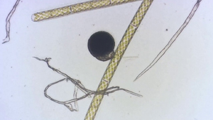
Time lapse: A hair was glued to a microscope slide and an amoeba was urged to walk over the hair. Each frame in the video comprises a stack of 10 images with selected overlapping depths of field. Note in the first half of the video how the camera view goes in and out of focus while all parts of the stacked view remain in focus. Details: Frame rate of capture for each stack = 60 Hz Time lapse between stacks = 4 sec, Total number of images = 220, Total time in video = 15 min, View width = 940 micron, Koehler illumination, 5X objective, Depth of field = approx 33 micron |
 |
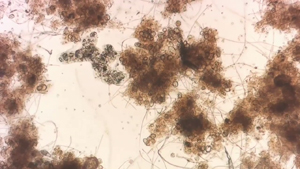
Time lapse: D. discoideum is commonly called a slime mold, but it is really a species of amoeba. In its life cycle, it exists as a collection of individual amoebae, a slug, and a fruiting body at the end of a stalk. This video shows motion, change of shape, and darkening of the fruiting body over a period of about 15 minutes. Each frame in the video comprises a stack of 10 images with selected overlapping depths of field. Note how the single-focus camera view goes in and out of focus while all parts of the stacked view remain in focus. Details: Frame rate of capture for each stack = 60 Hz, Time lapse between image stacks = 5 sec, View width = 940 micron, Episcopic illumination, 5X objective, Depth of field = approx 33 micron |
 |
This video shows slow-motion acquisition of 39 overlapping-DOF images between preset focal limits. The depth of field of the 50X objective lens of the microscope used was 1.18 microns; the thickness of the diatom collection is about 30 microns. The images in the video were actually taken at a rate of 60 Hz for a total acquisition time of all images of about 0.65 second. A 3D reconstruction obtained from the focus stacking operation is included at the end. |
 |
This video is a compilation of 515 images. Each frame is a focus stack of 26 images for a total of 13,390 images in the video. The depth of field of the 5X objective lens was 44 microns and the specimen depth was about 940 microns. The images in the video were taken at a rate of 60 Hz for a total acquisition time of 3.7 minutes. |
| GIFs: | |
 |
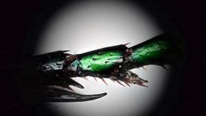
Dermestid larva anterior: This video shows the acquisition of 24 overlapping-DOF images of the anterior of a dermestid beetle larva that is emerging from the interior of a rat spinal column. The depth of field of the 5X microscope objective lens was 44 microns and the view of the beetle larva extends about 1016 microns into the spinal column. The images in this video were taken at a rate of 60 Hz for a total acquisition time of all images of about 0.4 seconds. |
 |
Dermestid larva posterior: This video shows the real-time acquisition of 60 overlapping-DOF images extending over the length of a dermestid beetle larva that is cleaning the inside of a rat spinal column. The depth of field of the 5X microscope objective lens was 44 microns and the beetle larva extends about 2540 microns into the spinal column. The images in this video were taken at a rate of 30 Hz for a total acquisition time of all images of about 2 seconds. |
 |
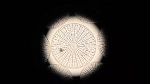
Arachnodiscus ehrenbergii: This video shows the real-time acquisition of 41 overlapping-DOF images between preset focal limits. The depth of field of the 20X objective lens of the microscope used was 2.8 microns; the thickness of the diatom collection is about 30 microns. The images in the video were taken at a rate of 60 Hz for a total acquisition time of all images of about 2/3 second. |
 |
Muscle-tendon junction: This video shows 4 overlapping-DOF images and a focus-stacked compilation of all 4. The thickness of the specimen is 7 microns, the depth of field of the 20X objective is 2.8 microns. The 4 images were taken at a rate of 60 Hz for a total image acquisition time of 0.07 sec. Note the improved focus in the final image. |
 |
Electronic test fixture: This video is a compilation of 17 images. The final image is a stack of 17 images taken from a video stream. The depth of field of the 5X objective lens was 44 microns and the specimen depth was about 1067 microns. The 17 images were taken at a rate of 60 Hz for a total image acquisition time of 0.28 sec. |